
Sensory receptive fields and synaptic contacts
In this semi-biographical piece, Raymond Noble revisits the work he produced for his Doctorate at the Royal Veterinary School, University of Edinburgh, in the late 1970s. He presents a scientific research article from The Journal of Physiology (1982) titled “Connexions between hair follicle afferent fibres and spinocervical tract neurones in the cat: the synthesis of receptive fields” by A. G. Brown and R. Noble.
Key Takeaways
- Raymond Noble’s article reflects on his doctoral research regarding sensory receptive fields and synaptic function.
- The study focused on how hair follicle sensory nerve fibres connect with spinocervical tract neurones, influencing sensory processing.
- Noble confirmed Ramón y Cajal’s theories on the relationship between neuronal structure and function, highlighting the significance of dendritic organisation.
- The research demonstrated how the arrangement of sensory inputs impacts synaptic connections and the processing of sensory information in the spinal cord.
- Noble’s findings reveal that proximal afferents form stronger synapses, affecting the overall sensory response in the nervous system.
Estimated reading time: 10 minutes
My doctoral work in the late 1970’s focused on mapping how cells formed a somatosensory representation of the skin’s surface in the spinal cord. I also measured how specific synapse locations on dendritic trees influenced cell function.
Our study examined how hair follicle sensory nerve fibres (afferents) connect with sensory neurones—specifically, spinocervical tract (s.c.t.) neurons—in the cat spinal cord. In particular, it investigated the organisation of their receptive fields and synaptic contacts. We hypothesised that afferents with receptive fields in the central region would form more synaptic connections near the sensory neurone’s cell body. By contrast, we expected afferents in the peripheral, outer region to form fewer synapses, located on dendrites farther from the soma.
The study showed how the spinal cord integrates sensory information from the skin, revealing that the spatial arrangement of receptive fields shapes the strength and location of synaptic connections. This has broader implications for understanding sensory processing and neural circuit organisation in mammals.
Ramon Y Cajal – the architecture of neural networks
During my PhD, I often referred to Ramón y Cajal’s work. He won the Nobel Prize for his research on the structure of the nervous system. He revealed the fine architecture of sensory nerve endings and proposed remarkable hypotheses about their functions. Much of my PhD research confirmed his assumptions. I spent much of my time studying his detailed drawings of neurone structure and how their anatomical relationships shaped the central nervous system. Cajal’s work was highly detailed and cross-species, but at that time he could only speculate about the specific functions of the neurones he described.
I read the French edition of Cajal’s book, which gives different neuron types beautiful floral names based on the shapes of their arbors and axon branches. Dendrites, which resemble the branches of trees or bushes, receive functional connections—called synapses—from other neurons. Accordingly, “neurones de double bouquets” describes neurons with two distinct arbors arranged in columns. This terminology highlights how these neurons connect with others, whether by receiving input or sending it onward. Through meticulous study, Cajal revealed the central nervous system’s intricate architecture and offered insights that make his work especially compelling.
How Neurons Process Sensory Input
Neurons form intricate, tree-like structures, with branches called dendrites arranged in varied patterns. These dendrites receive thousands of synaptic terminals from other neurons, creating functional networks. Our task was to understand how a neuron processes this abundance of synapses to produce meaningful outputs. In this case, sensory receptors in the skin provided the input. The skin contains many different types of touch receptors. We identified how each type had distinctive arborisations in the spinal cord.
In 1909, Cajal proposed that this anatomy had functional significance. Decades later, researchers confirmed the importance of dendrites and axon branches in the cerebral cortex. For example, in the 1960s, Hubel and Wiesel showed that cells in the visual cortex, arranged in columns, responded to specific angles of light. Moreover, the preferred angle shifted from one column to the next, showing how the cortex’s columnar organisation aligned with its function.
My thesis showed how the neuronal architecture relates to cutaneous sensory function at the first relay in the spinal cord.
Organisation of somatosensory networks
I joined Alan Brown’s research group in Edinburgh in 1976 to study how somatosensory neuronal networks are functionally organised. We used a technique to stain the axons, branches, collaterals, cell bodies, and dendrites of functionally identified neurons.
My doctoral work focused on mapping how cells formed a somatosensory representation of the skin’s surface in the spinal cord. I also measured how specific synapse locations on dendritic trees influenced cell function.
Hair-follicle sensory receptors
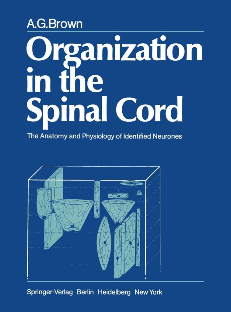
One group of arborisations in the dorsal horn of the spinal cord particularly caught our attention. By identifying specific axons and neurons through their physiological activity, we could label them for later histological analysis. We found that the arborisations Cajal described originated from sensory receptors in the skin, including hair follicles and vibration-sensitive Pacinian corpuscles. Moreover, each type of skin receptor forms a distinct pattern of terminal arborisation in the spinal cord (see Alan G. Brown’s book). Organization in the Spinal Cord: The Anatomy and Physiology of Identified Neurones)
A hair follicle is a tiny, tube-like structure in the skin that surrounds the hair root and produces the hair strand. It also serves as a sensory organ: nerve endings within the follicle detect hair movement. We inserted a glass-capillary microelectrode filled with electrolyte and an enzyme into the axon as it entered the spinal cord. This allowed us to map the axon’s location and receptive field on the skin. Subsequent histology revealed axonal arborisation and synaptic boutons in the spinal cord for further analysis.
Linking Sensory Function and Spinal Cord Anatomy
Nearly seven decades after Ramón y Cajal used Golgi staining, we confirmed Cajal’s suggestion. The arborisations he observed belonged to specific sensory fibres entering the spinal cord from the skin. Cajal could not test this hypothesis. We recorded and labelled these fibres, advancing our understanding of the relationship between function and anatomy in the somatosensory system. Furthermore, each type of skin sensory organ gives rise to a distinct, characteristic arborisation pattern in the spinal cord.
Longitudinal Organization of Sensory Inputs
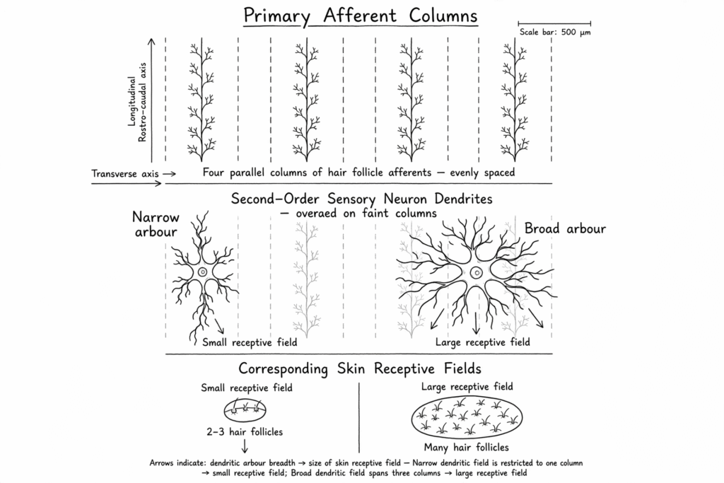
Hair follicle afferent arborisations formed continuous columns along sections of the spinal cord. Sensory neuron dendrites received synapses from these columns and generally ran longitudinally alongside the afferents. However, some neurons had larger dendritic arbours that extended across multiple afferent arborisations. These broader arbours tended to correspond to larger receptive fields, highlighting a link between dendritic extent and sensory field size.
The Synthesis of Receptive Fields
Sensory axon collaterals form overlapping columns in the dorsal horn, enabling second-order sensory cells to recruit them systematically. The schematic illustrates how the columnar arrangement of primary sensory fibres and their axon collaterals relates to the arrangement of second-order sensory neurons and receptive field size. At the top, primary sensory axon collaterals form overlapping longitudinal columns. In the middle panel, the shape and size of the sensory neurons allow them to form synapses with these columns. For simplicity, the diagram shows the arborisations evenly spaced. The numbers are for illustrative purposes only.
Intracellular recording of identified neurones
For neuroscientists in the 1970s, recording from inside a neuron was the ultimate achievement. The work was challenging and exhilarating. We used a delicate glass microelectrode—a tiny, electrolyte-filled capillary tube. After heating and pulling it to form a microscopic tip, we used the electrode to penetrate the cell membrane. Our recordings captured membrane potentials, small voltage fluctuations, and action potentials—the distinct electrical signals neurons carry along their axons. Every detail was documented in our lab notebooks. Our vivid descriptions often conveyed the thrill of finding the neuron we sought. This painstaking work made neurons’ hidden electrical activity directly observable.
Additionally, we could label the recorded cell for later histological identification. This technique allowed us to label the cell body, dendrites, and axon.
Intracellular recordings
Searching is the operative word: we first identified the neuron by mapping its receptive field’s location, modality, and size. Then, by moving the microelectrode systematically, we homed in on the neuron in the spinal cord. Once the electrode touched the cell membrane, we nudged it down a tiny fraction and penetrated the membrane. The sudden drop in potential—to about −50 to −70 mV—confirmed that we were inside. Next, we let the cell settle after the disturbance. Soon, we could see—and hear—the membrane potentials’ soft murmur, punctuated by the occasional action potential. Finally, by gently brushing the skin’s hairs, we mapped the receptive field.
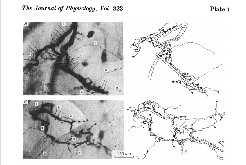
But then came the pièce de résistance. We passed an electrical current through the electrode, carrying the enzyme horseradish peroxidase (HRP) into the cell. The HRP then travelled through the dendrites and axon collaterals. In histology, we added a reagent that reacted with HRP to produce a stain revealing these processes. This enabled us to identify and trace the single sensory axon. This revealed the synapses it formed with the dendritic tree of the identified sensory neuron (Figure 1).
Imagine how difficult it was: the sensory nerve fibres entering the spinal cord were only a few micrometres in diameter, and we had to place a fine-tipped electrode inside them. The work was painstaking at times. However, the reward was significant.
Comparison of Central vs Peripheral Contacts
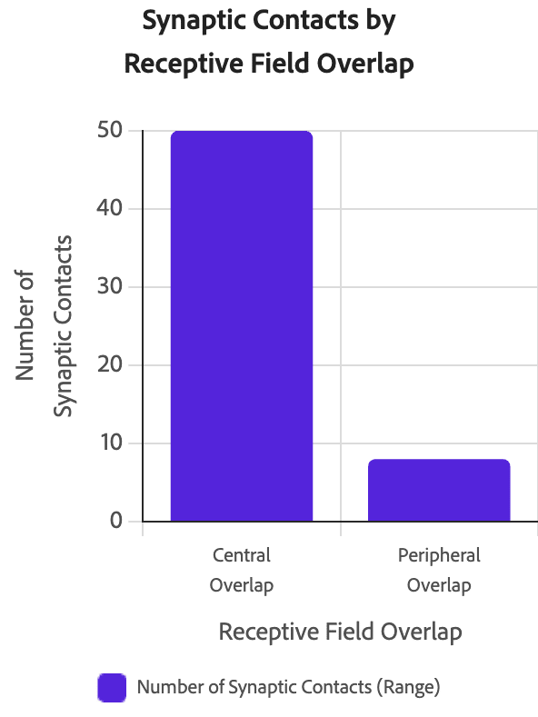
Our study showed that hair-follicle receptors with receptive fields centred within the sensory neuron’s receptive field formed more synapses. These synapses were closer to the cell body. By contrast, receptors at the periphery formed fewer synapses, located on distal dendrites (Figure 3).
Figure 1 shows the synaptic contacts between a single afferent fibre and a somatosensory cell in the spinal cord. The afferent field is central to the somatosensory cell’s receptive field. The micrographs on the left show the labelled dendrites, afferent fibre, and synaptic boutons.
First stage processing can be complex
This comparison shows how receptive field relationships shape the anatomical and functional nature of synaptic connections in sensory pathways. The central region of the receptive field makes a strong monosynaptic connection. Later work showed that even a single impulse in the hair-follicle axon triggered an amplified response in the somatosensory cell. However, inhibitory interneurons also modified this response.